Posts

Showing posts from March, 2020

RNAi Mechanism : RNA interference phenomena in cell

Introduction: Here is an amazing topic in our RNA science and technology that is RNA interference and gene silencing . RNAi mechanism: The whole phenomena is performed inside a cell which targets to silence mRNA Strand and break it down . Where this function is found ?  The function of RNA interference is found in both  Prokaryotic and  eukaryotic cells . # The discoverer of RNA interference is Andrew Fine  and Craig Mello . Here we just briefly study the basic function and future application of RNA interference. It has a massive potential to make a lots of changes in human life. Lets begin .... The basically the RNA interference actual goal is to hamper the protein synthesis . We know RNA is single stranded but any how or by any chance Virus produce a double stranded RNA it could be  very specific target side to produce RNAi . It leads to produce Si RNA ,Sh RNA ,Mi RNA. Here Dicer protein helps trim dsRna to form Si RNA ,Mi RNa ,Sh RN
Microtubules motors and movement

Image
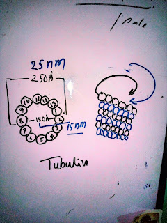
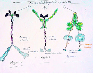
Introduction: Microtubules are responsible for different kinds of cell movements including the intracellular transport and positioning of membrane vesicles and organelles, the separation of chromosomes at mitosis and the beating of cilia and flagella .Motor protein's action and the polymerization and depolarization of microtubules use the energy from ATP hydrolysis to regulate the movement and force . There are two large families of motor protein which mainly responsible for powering of movements where the microtubules participate. Kinesin 1 motor protein and Dynein : There  are two major class of motor protein associated with movement along microtubules. 1.kinesin 1:This  is a  protein belonging to class of motor protein found in eukaryotes.  Kinesin 1 was firstly discovered kinesin. Kinesin is like a vehicle which move along the microtubules filament by hydrolysis of  ATP ,has some cellular functions like mitosis, meiosis and transport of cellular cargo .Kinesin walks

The journey to the discovering of DNA : A molecule changed the world

Abstract: If I ask you ,what is DNA ...? You may reply that it is a hereditary material found in almost every organism,contains the genetic instructions of organism for its development or functions...etc. But today you could learn a history of DNA .  I just tell you to pack your excitement to the top and let's know something new.  The Discovery of DNA : There are so many people who had already guess that the discovery of the DNA was done by Watson and Francis Crick but it is fully wrong . In 1860s ,a Swiss chemist ,Johann Friedrich Miescher unknowingly discovered DNA . One day Johann planned to research on white blood cells' key components .He collected pus coated bandages from a nearby medical clinic . Johann use salt solution to understand more about WBC and further he  added acid to the solution . Then a substance separated from the solution. Next he added alkali and it dissolved again .However during this process he understood that the substance has an unique

The Cell Membrane: Movement of phospholipids in Lipid bilayer

Image
Abstract : Phospholipid is a major component which show different types of movements. Here we will describe 5 types of movement of lipid in lipid bilayer. So don't be late ..       Lets learn.... https://pin.it/2J6aICm Lateral diffusion Rotation Swing Flexion 5.Flip-Flop/Transverse diffusion.        As a component we took phospholipid to show you the procedure very easy way . #1 : Lateral diffusion : Cell membrane molecules transpose with it's local molecules . Lateral movement also provide cell membrane fluid structure. It has been discovered that phospholipid moves via hop diffusion. #2:  Rotation: It occurs when a lipid molecule rotates itself very fast around it's axis and interact with intermidiate neighbor molecules. #3:   Swing: Molecules swing from one side to another one. #4: Flexion : The movement is started by contraction.  #5: Flip-Flop movement/Transverse movement: However Flip-flop movement and transverse diffusion is

Don't make panic and stop spreading rumors ## Hantavirus

PLEASE DON'T MAKE PANIC ABOUT HANTAVIRUS -IF U GONNA EAT RATS ...  Hantavirus is a known virus and not a new one .You are wrong if you think that the virus is communicable human to human . It does not get transmitted through human to human. So how they spread????  Various rodents are get affected by this virus ,when people come contact them or an area of their faces and urine present ,that will be a good chance for the virus to enter human through the air inhaled .  According to CDC the correct incubation period is not known properly. Symptoms: Fever,fatigue, headaches ,even vomiting and abdominal pains are also indicate its symptoms . According CDC anyone who comes contact with this virus by rodents has a risk of Hantavirus pulmonary syndrome. Treatments: There is no absolute vaccines are available .But they can be killed by such disinfectant such bleach , detergents and alcohol . So don't make panic and stay strong.        Thank you ..... Visit my websi

Overview of cytoskeleton

Image
Highlights: #What is Cytoskeleton? ## what are the composition of Cytoskeleton? ## Functions ... Abstract: Today we will discuss about the above mentioned matter. By seeing my post you will know overall about cytoskeleton .  Before study in molecular level you should have to know an overview of cytoskeleton .  Lets start our journey .... # Cytoskeleton is a dynamic structure which provide structural framework for cell ,serve itself as a scaffold that fix cell shape and determine the position of cytoplasm. It is responsible for movement of cell. Next discuss about major  Component of cytoskeleton .. ## There are three types of major or principle component of cytoskeleton . 1.Actin Filaments  2.Microtubules 3.Intermediate Filaments ### Lets try to understand their structure and way of function briefly. Actin Filaments : It also known as Microfilament .They are produced by one molecule is actin . It is fine thread like structure of protei

Vesicle formation of Cop I and Cop II

Image
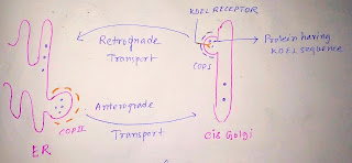
Learn how COP I and COP II vesicle formed in diagram in step by step. Abstract: It is very interesting to know how COP I & COP II vesicles formed when Retrograde and Anterograde transport occur.... Today we will highlight on the mentioned matter. Lets go.. #An imaginary picture of anterograde and retrograde. Endoplasmic Reticulum resident protein has KDEL sequence at the protein c terminal end . KDEL receptor present in Golgi. KDEL receptor recognise ER resident protein and transport the protein to ER (Retrograde transport) by COP I . ER transport their protein by vesicular transport by using COP II to Golgi. Formation of COP I coated vesicle: 1.Recruitment of Sec7 (GEF) . 2. After recruitment of ARF1 from cytosol, Sec7  converts GDP to GTP of ARF1 and make it active from inactive form. 3.Hydrophobic tail present in ARF1 GTP which inserted into golgi membrane.  4.Finally COP I is activated by ARF1 . 5.As the picture before fusion the v

Mechanism of Vesicles Docking and Fusion

Image
Abstract:  Analysis of Biochemical and Genetics study reveal a molecular mechanism which is selectively cargo molecule transport between donor and target compartments .                                        Different membrane proteins participate in vesicular fusion .         #We will discuss about some membrane proteins which participate in vesicles docking and fusion . Protein involved in vesicle docking 1: SNAREs protein: Complimentary sets of vesicles SNAREs(v-SNARE) and target membrane (t-SNARE) contribute to the selectivity of transport vesicle docking and fusion.   v-SNARE: During the budding of transport vesicles it is packaged together with coat protein from donor membrane and then bind to complementary t-SNARE in target membrane.   t-SNARE: After the fusion t-SNARE Mechanism and v-SNARE make tight complex     Next this complex have to dissociate so that the  SNARE protein( v-SNARE) can be recycled to the donor compartment for d